AoMP-001-Q: Classic Artifacts in Radial MRI and a Mysterious Artifact (re-discovered)
A deep dive into understanding the physics behind classic radial MRI artifacts, a mysterious artifact associated with iterative reconstruction algorithms, and how to mitigate them.
Table of Contents
“Mystery creates wonder and wonder is the basis of man’s desire to understand.”
-- Neil Armstrong (1930–2012)
This is the first challenge in the AoMP series, in which I present a set of artifacts that I stumbled upon during my research.
TL;DR: In this challenge, you will learn about the classic artifacts in radial MRI. You will also encounter a so-called “mysterious” artifact. By solving this challenge, you will gain deep intuition about the physics behind these artifacts and how to mitigate them. The knowledge and intuition you gain are not only useful for radial MRI, but also for understanding MRI physics, other MRI artifacts, and signal processing and engineering more broadly.
If you have any suggestions or feedback, I would love to hear from you.
Radial MRI
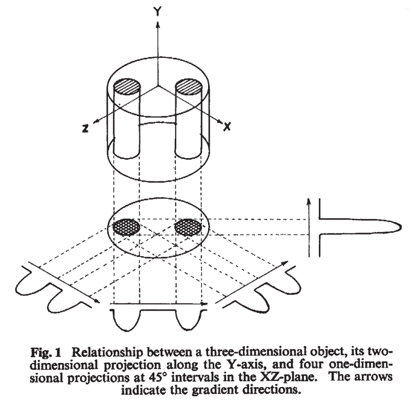
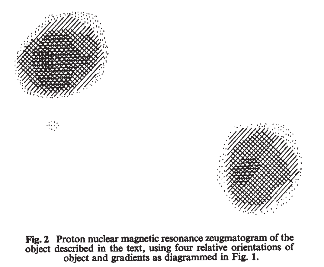
Did you know that the first MRI image, acquired in 1973 by Paul Lauterbur, was actually radial? This pioneering work contributed to his receiving the Nobel Prize in Physiology or Medicine in 2003.
Although used to create the first ever MRI image, the radial MRI acquisition scheme is not commonly used in clinical practice today. Instead, the Cartesian acquisition scheme is more widely adopted in clinical MRI due to its simplicity, efficiency, and robustness to system imperfections. However, radial MRI has unique advantages such as reduced motion artifacts and applicability in certain applications, which has led to its continued use in research and specialized clinical settings.
With the improvement in hardware and reconstruction algorithms, radial MRI has been gaining more attention in recent years for its potential to provide high-quality images with reduced artifacts, especially in applications such as free-breathing, free-running 3D/4D/5D cardiac imaging and liver imaging. Radial MRI is perfectly suited for these applications because of its inherent robustness to motion artifacts as well as its ability to provide navigation information for motion correction and reconstruction.
Clinically, radial MRI has been used in liver MRI to reduce motion artifacts and capture the dynamic contrast enhancement of liver lesions. It also has been used in musculoskeletal MRI to generate CT-like contrast via ultrashort echo time (UTE) and zero echo time (ZTE) imaging.
Although not strictly radial, the PROPELLER/BLADE/JET MRI acquisition scheme, which samples k-space in a rotating fashion, has been widely used in brain and body MRI to reduce motion artifacts, where the artifacts are similar to those in radial MRI.
In this post, we will challenge you to identify the causes of the classic artifacts in radial MRI and propose solutions to mitigate them. During the process, we will also re-discover a mysterious artifact and strategy to mitigate it.
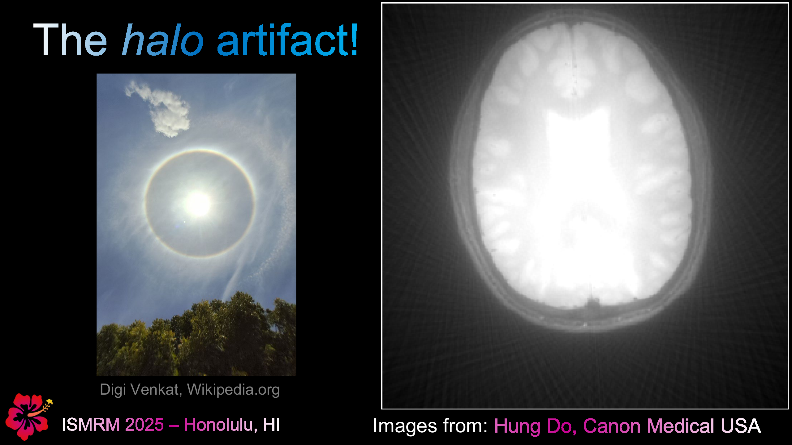
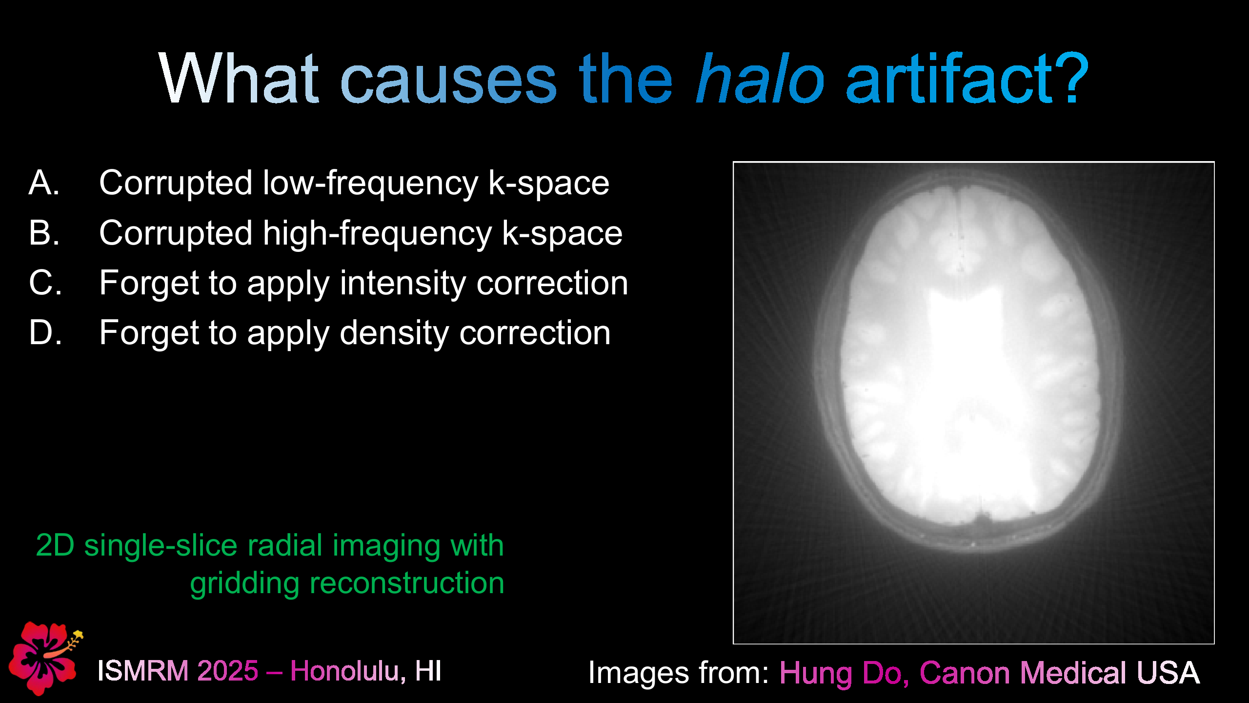
The “Halo” Artifact
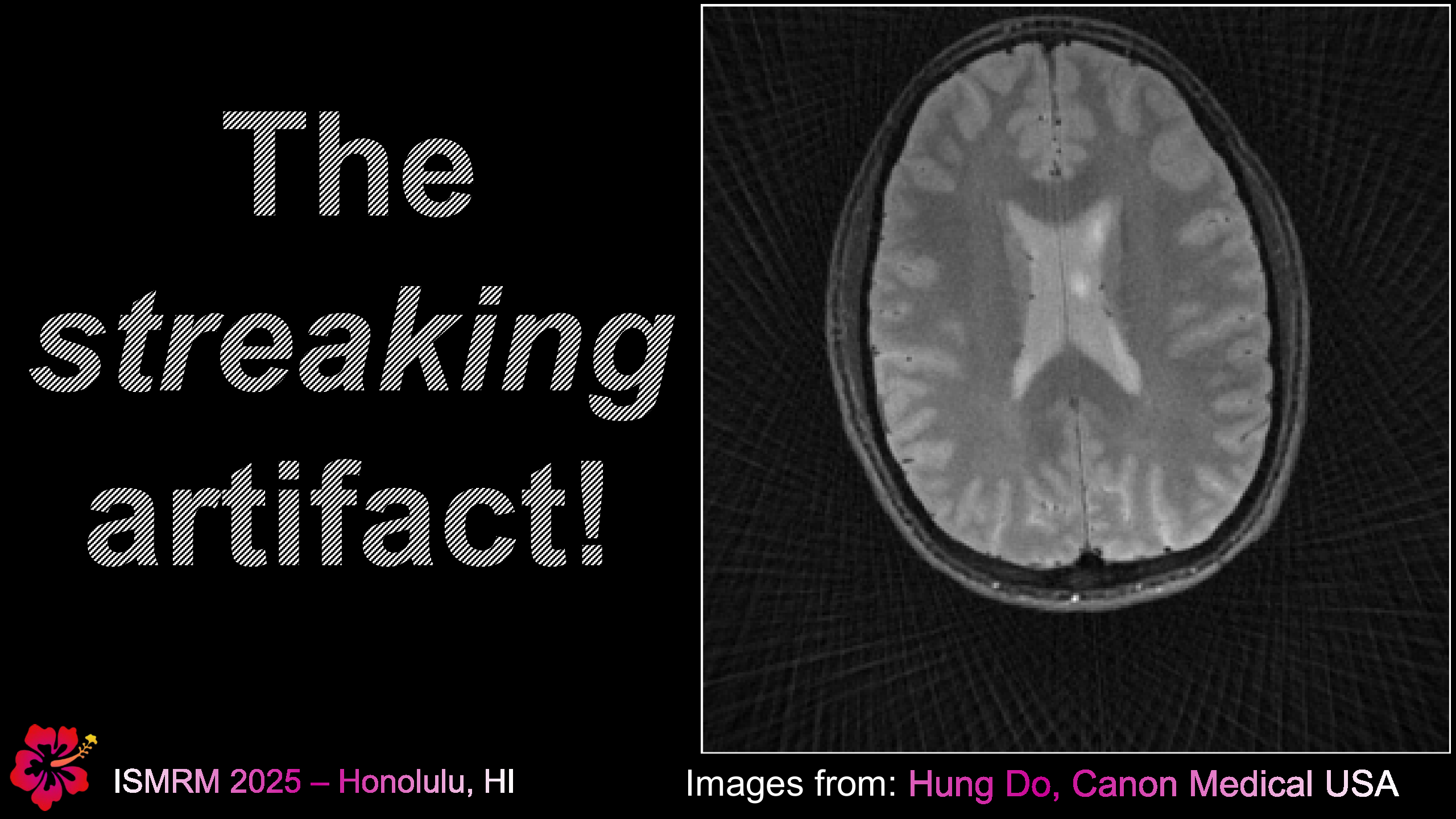
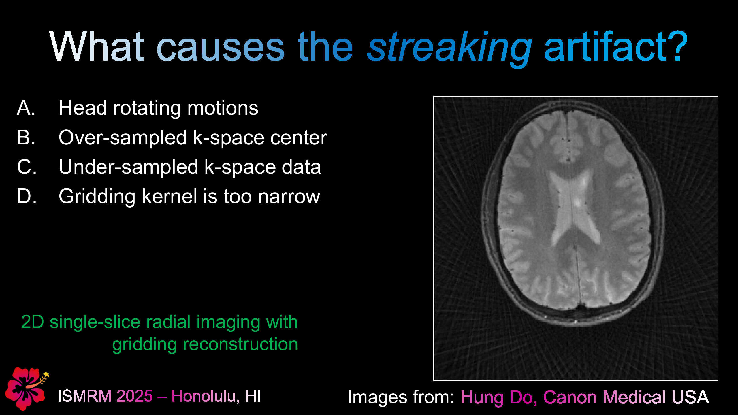
The “Freaking” Artifact
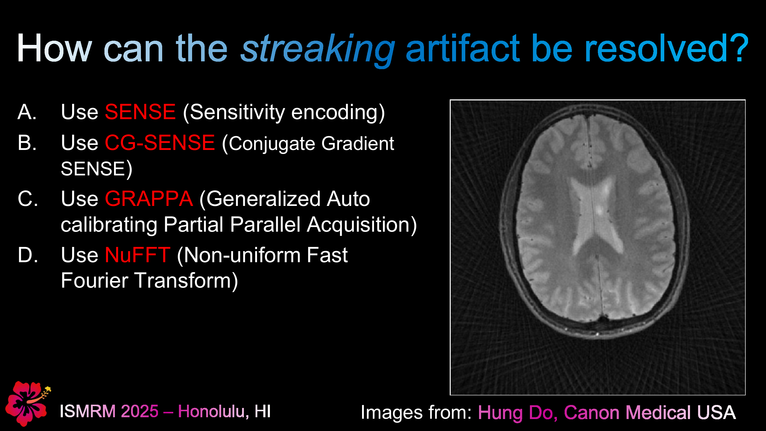
How to Mitigate the “Freaking” Artifact?
The “Mysterious” Artifact
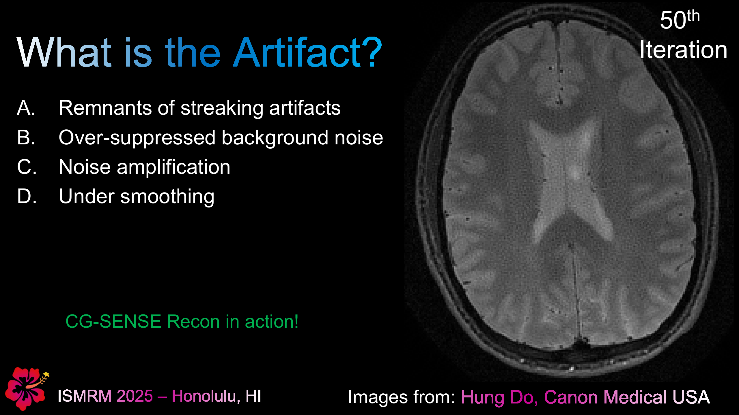
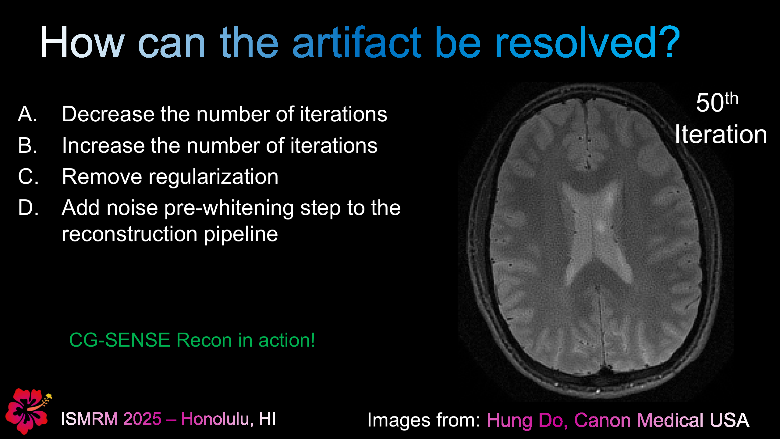
How to Mitigate the “Mysterious” Artifact?
Citing this work
If you found this useful, please cite this as:Hung P. Do, PhD, MSEE (Feb 2026). “AoMP-001-Q: Classic Artifacts in Radial MRI and a Mysterious Artifact (re-discovered)”. The Art of MRI physics (AoMP). https://hdocmsu.github.io/blog/2026/02/27/aomp-001-q/
or as a BibTeX entry:
@article{do-20260227-aomp-001-q,
  title   = {AoMP-001-Q: Classic Artifacts in Radial MRI and a Mysterious Artifact (re-discovered)},
  author  = {Do, Hung P. PhD, MSEE},
  journal = {The Art of MRI Physics (AoMP)},
  year    = {2026},
  month   = {Feb},
  url     = {https://hdocmsu.github.io/blog/2026/02/27/aomp-001-q/}
}
Get future posts delivered to your inbox.